Shoulder Tendon And Ligament Anatomy : Shoulder Pain And Problems Johns Hopkins Medicine : At the level of the pip joint, the.. Anatomy of the canine shoulder (scapula, humerus, ligaments, shoulder joint, muscles and tendons) on ct. Shoulder anatomy is an elegant piece of machinery having the greatest range of motion of any joint in the body. There are several important ligaments in the shoulder. (3) a syndesmosis is a joint in which a ligament connects two bones, allowing for a little movement (amphiarthroses). A joint capsule is a watertight sac that surrounds a joint.
More about dental anatomy and periodontal ligaments you can find in the article about the anatomy of the teeth and this interesting video tutorial. Muscles, tendons, and ligaments run along the surfaces of the feet, allowing the complex movements needed for motion and balance. The capsule, extensor tendon, and skin are very thin and lax dorsally, allowing for both phalanx bones to flex more. Superior glenohumeral ligament and coracohumeral ligament are the primary restraints to posterior translation with the are flexed, adducted and prevents inferior translation and external rotation in the abducted shoulder, and provides stability to the long head of the biceps tendon (neer cs ii, corr. However, many tendon and ligament injuries can be avoided through proper conditioning and training regimens and by not pushing a horse beyond its limits in racing or other competitions.
Humeral Articulation Or Shoulder Joint Human Anatomy from theodora.com The joint, held in place by a ligaments, tendons, and muscles, behaves in a unique manner allowing a large range of motion of the arms. Tendons and ligaments are bands of connective tissue that help stabilize the body and allow movement. Ligaments aid in joint stability during rest and movement and help prevent injury from hyperextension and hyperflexion (excessive movements). Anatomy of the canine shoulder (scapula, humerus, ligaments, shoulder joint, muscles and tendons) on ct. In addition to the bones and joints, the shoulder contains a network of soft tissues, such as muscles, tendons, and ligaments. Learn about the muscles, tendons, bones, and ligaments that comprise the knee joint anatomy. • now that we've taken a look at the bones, joints and ligaments, let's spend about a minute reviewing what we've seen so far. These ligaments are main source of stability for the shoulder.
Shoulder joint allows lifting, pushing and pulling by upper extremity.
Tendons and ligaments commonly sustain injuries, which usually have similar symptoms and treatments. Muscles, tendons, and ligaments run along the surfaces of the feet, allowing the complex movements needed for motion and balance. Learn about the muscles, tendons, bones, and ligaments that comprise the knee joint anatomy. It is a complex structure of bones, muscles and ligaments with the ability to lift weights and create enormous strength. The anatomy of the provides the strength and functionality of the upper body. In addition to the bones and joints, the shoulder contains a network of soft tissues, such as muscles, tendons, and ligaments. Once the ligaments, tendons, and muscles around the shoulder become loose or torn, dislocations can occur repeatedly. In the horse, lateral and medial movements of this joint are impossible due to the shape of the humeral head; Shoulder joint is formed by a group of ligaments that connect humerus to glenoid. The shoulder can counteract an extreme impact but is also vulnerable to to a range of pathologies due to. The distal joint between the tibia and fibula is an example of a. Anatomy of the shoulder these pictures of this page are about:shoulder tendons and ligaments anatomy. The shoulder joint is the articulation between the glenoid cavity of the scapula and the head of the humerus.
Anatomy of the canine shoulder (scapula, humerus, ligaments, shoulder joint, muscles and tendons) on ct. Anatomy of the human body via wikimedia commons, public domain. Video atlas of human anatomy. Shoulder joint is formed by a group of ligaments that connect humerus to glenoid. The anatomy of the provides the strength and functionality of the upper body.
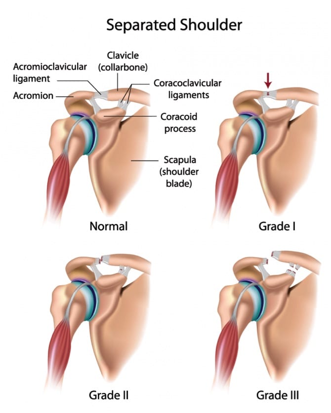
3 Grades Of Separated Shoulder Diagnosis And Treatment from charmaustin.com Shoulder ligaments and tendons diagram quizlet from o.quizlet.com. Anatomy of the canine shoulder (scapula, humerus, ligaments, shoulder joint, muscles and tendons) on ct. Shoulder ligaments at louisiana state university. The distal joint between the tibia and fibula is an example of a. These ligaments are main source of stability for the shoulder. Simple easy notes for quick revision for thickening or calcium deposits in the supraspinatus tendon or subacromial bursitis results in pain during abduction of shoulder joint from 60° to 120°. This instability is countered by the strength of the rotator cuff muscles, tendons, ligaments, and the glenoid labrum. Shoulder joint allows lifting, pushing and pulling by upper extremity.
It is a complex structure of bones, muscles and ligaments with the ability to lift weights and create enormous strength.
These imaging studies create better pictures of soft tissues. 178 просмотров • 14 апр. The achilles tendon connects the heel to the calf muscle and is essential for running, jumping, and standing on the toes. Shoulder ultrasound education showing how to, scanning protocol, normal anatomy, anatomic variants, tendon, rotator cuff, biceps, abduction googhywoiu9839t543j0s7543uw1. Anatomy of the shoulder these pictures of this page are about:shoulder tendons and ligaments anatomy. Anteriorly the subscapularis tendon is separated from the supraspinatus tendon by a gap, the rotator interval, which these ligaments pass from the coracoid and glenoid respectively, and insert into the humeral head on either. Movement is therefore limited to flexion and extension. Tendons and ligaments commonly sustain injuries, which usually have similar symptoms and treatments. These ligaments are main source of stability for the shoulder. Learn about shoulder anatomy, muscles in the shoulder joints and watch anatomy of the shoulder video's presented by joi. The distal joint between the tibia and fibula is an example of a. Start studying shoulder ligaments and tendons. Learn vocabulary, terms and more with flashcards, games and other study tools.
Video atlas of human anatomy. Anatomy of the shoulder these pictures of this page are about:shoulder tendons and ligaments anatomy. The anatomy of the provides the strength and functionality of the upper body. Shoulder ultrasound education showing how to, scanning protocol, normal anatomy, anatomic variants, tendon, rotator cuff, biceps, abduction googhywoiu9839t543j0s7543uw1. Tendons, ligaments, bone, and cartilage are connective tissues in which the activities of various cellular populations are responsible for synthesis and maintenance of large amounts of extracellular matrix that should, theoretically, be dynamically optimized to respond to mechanical demands.
Shoulder Joint Anatomy Physiology Movement Exercise from samarpanphysioclinic.com Mri may help your doctor identify injuries to the ligaments and tendons surrounding your shoulder joint. (3) a syndesmosis is a joint in which a ligament connects two bones, allowing for a little movement (amphiarthroses). The achilles tendon connects the heel to the calf muscle and is essential for running, jumping, and standing on the toes. Tendons and ligaments commonly sustain injuries, which usually have similar symptoms and treatments. Learn about the muscles, tendons, bones, and ligaments that comprise the knee joint anatomy. However, many tendon and ligament injuries can be avoided through proper conditioning and training regimens and by not pushing a horse beyond its limits in racing or other competitions. The distal joint between the tibia and fibula is an example of a. Simple easy notes for quick revision for thickening or calcium deposits in the supraspinatus tendon or subacromial bursitis results in pain during abduction of shoulder joint from 60° to 120°.
The shoulder joint is the articulation between the glenoid cavity of the scapula and the head of the humerus.
Shoulder joint allows lifting, pushing and pulling by upper extremity. Ligaments and tendons are fibrous bands of connective tissue that attach to bone connecting two or more bones together and help stabilize joints. More about dental anatomy and periodontal ligaments you can find in the article about the anatomy of the teeth and this interesting video tutorial. These ligaments are main source of stability for the shoulder. Tendons, ligaments, bone, and cartilage are connective tissues in which the activities of various cellular populations are responsible for synthesis and maintenance of large amounts of extracellular matrix that should, theoretically, be dynamically optimized to respond to mechanical demands. A joint capsule is a watertight sac that surrounds a joint. • now that we've taken a look at the bones, joints and ligaments, let's spend about a minute reviewing what we've seen so far. Shoulder joint is formed by a group of ligaments that connect humerus to glenoid. Shoulder tendonitis is inflammation of your rotator cuff or bicep tendons, often caused by overuse of. Patellar tendon problems can arise from knee arthritis but are more likely to affect athletes who do a lot of running, pivoting, and jumping. Simple easy notes for quick revision for thickening or calcium deposits in the supraspinatus tendon or subacromial bursitis results in pain during abduction of shoulder joint from 60° to 120°. 178 просмотров • 14 апр. Anatomy of the canine shoulder (scapula, humerus, ligaments, shoulder joint, muscles and tendons) on ct.
Start studying shoulder ligaments and tendons shoulder tendon anatomy. The shoulder joint (glenohumeral joint) is a ball and socket joint between the scapula and the humerus.


0 Komentar